
- Date:
- January 26 (Mon.) -28 (Wed.), 2015
- Venue:
- Kyoto International Conference Center
Report
This symposium concluded the activities associated with the grant on “Multi-dimensional Fluorescence Live Imaging of Cellular Function and Molecular Activities,” by the Ministry of Education, Culture, Sports, Science, and Technology, Japan. The symposium also aims to provide a venue where researchers engaged in fluorescence live imaging can exchange their expertise, and a forum for heralding new grant projects for the coming years. More than 300 hundred attendees have enjoyed the exciting discussions and cutting-edge presentations at this event. *When a cursor is put on the image, the expansion display is done. Moreover, it opens in another window when clicking.
Plenary lecture
Dr. Eric Betzig
The first plenary lecture was given by Dr. Eric Betzig, who won the Nobel Prize in Chemistry in 2014. Surprisingly, he talked only slightly about PALM (Photoactivated localization microscopy), the invention which brought him the Nobel Prize, but focused his fascinating talk on the development and the power of SIM (Structured Illumination Microscopy). By using Bessel beam illumination, he succeeded in reducing phototoxicity and increasing spatial resolution beyond the diffraction limit. The videos shown in his talk were simply remarkable. Cells with waving lamellipodia and filopodia were visualized at the resolution that we could achieve only saw by the scanning electron microscope.
Dr. Lihong Wang
The second plenary lecture was given by Dr. Lihong Wang, who is leading the development of photoacoustic tomography (PAT). He overviewed the current progress of PAT and surprised the audience with his introduction of superresolution PAT, which brings the resolution of PAT close to that of optical microscopy, and the imaging depth beyond the width of the usual lab mice.
These two lecturers reminded us of the old adage, “Records are made be broken!”
Session 1
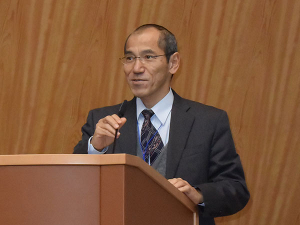
Atsushi Miyawaki chaired Session 1, “Cutting Edge Technology of Fluorescence Imaging.” Atsushi Miyawaki presented a number of fluorescent proteins and probes that he developed. Among them, UnaG isolated from Japanese eels was found to be a useful tool to develop a very sensitive assay system for bilirubin. He talked about his dream of applying this assay to diagnose neonatal jaundice. Samie Jaffrey talked about the application of RNA aptamer technology to create a fluorescent RNA known as Spinach. Na Ji introduced applications of adaptive optics, which have been extensively developed for the acquisition of sharp images of stars in modern telescopes. So, by various interdisciplinary approaches, both microscopes and the fluorescent probes are rapidly progressing to open new fields of biology.
Atsushi Miyawaki
Samie Jaffrey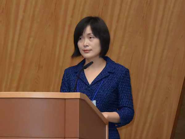
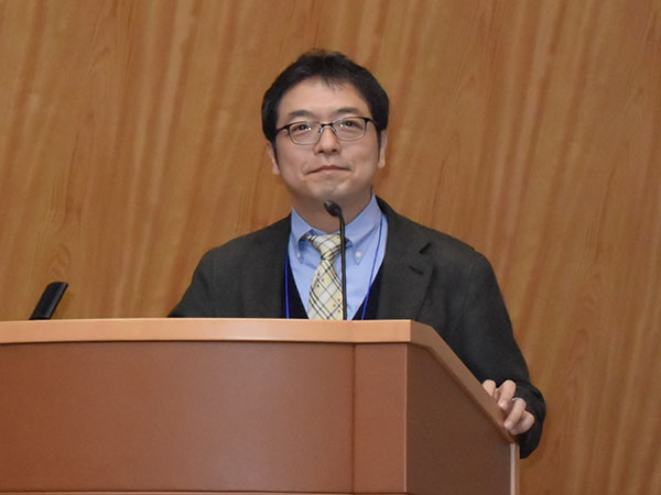
Na Ji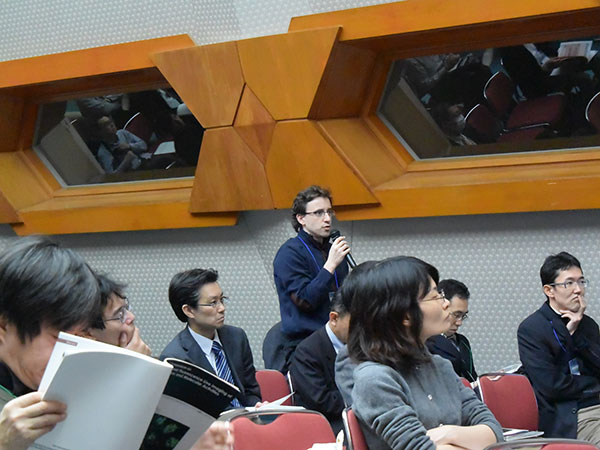
audience
Session 2
Tomomi Nemoto chaired Session 2, “Visualization and Optical Control of Neural Activity.” Tomomi Nemoto followed Na Ji’s talk on adaptive optics and showed that he had succeeded in visualizing the mouse brain to a depth of as much as 1.4 mm, which was sufficient to see the hippocampus from the surface of the cerebral cortex. Vatentin Nägerl gave a talk on another super-resolution technology named Stimulated Emission Depletion Microscopy, or STED. With his inventive 3D STED, he visualized how the structure of the synapse changes during memory formation. Masanori Matsuzaki presented his data on motor learning based on the calcium imaging of living mouse brains. These talks showed that fluorescent imaging enables us not only to see the structural but also the functional basis of learning and memory formation in the living mouse brain.
Tomomi Nemoto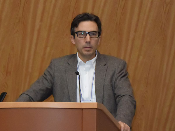
Vatentin Nägerl
Masanori Matsuzaki
audience
Session 3
On the second day, Takaharu Okada opened Session 3, “In Vivo Imaging by Multi-Photon Microscopy (MPE).” His beautiful two-photon videos, in combination with various genetically modified mouse strains, revealed how dendritic cells transmit signals to follicular B lymphocytes. Guy Shakhar also used two-photon microscopes to visualize how cytotoxic T lymphocytes (CTL) attack melanoma cells and to show the role of tissue oxygen concentration on the cytotoxicity of CTL. Kenji Kabashima studied contact dermatitis using MPE. He traced how dendritic cells in the skin lesion activate lymphocytes in the skin and showed the role of the CXCL2 signaling cascade in this phenomenon. Tatsuko Kinashi studied the role of Rap1 small GTPase on the integrin activation and T cell migration. He also showed the cross-talk between the Rap1 signaling cascade and the Hippo pathway. All four speakers in this session showed by MPE how vividly the immune cells move around and function in living tissues.
Takaharu Okada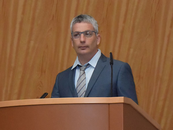
Guy Shakhar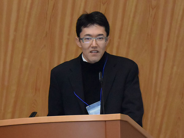
Kenji Kabashima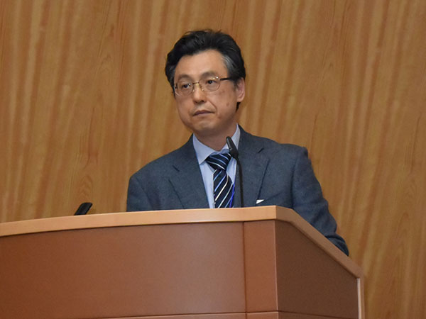
Tatsuko Kinashi
Session 4
Takeshi Imamura chaired Session 4, “Fluorescence Imaging of Cancer.” This session was also dedicated to MPE. Takeshi Imamura showed how tumor cells behave in living tissues. The AO technique was used again to clear the blurred image of cancer cells. Charles Lin met the challenge of imaging cells in the bone marrow. Using an oxygen nanosensor, he was also able to visualize the gradient of oxygen concentration for the first time. Michio Tomura used KikGR mice, in which the green color of KikGR fluorescent protein can be photoconverted to red, thereby enabling the tracking of macrophages. He showed elegantly how macrophage/dendritic cells bring tumor antigens to the regional lymph nodes by this technique.
Takeshi Imamura
Charles Lin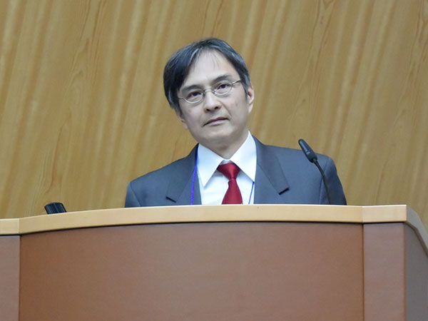
Michio Tomura
audience
Session 5
Session 5, “Visualization of Bone and Immune Systems,” was chaired by Masaru Ishii and dedicated to bone biology. Using MPE, Masaru Ishii visualized the dynamics of osteoblasts and osteoclasts in the bone marrow and their regulation by sphingosine-1-phosphate. He also presented the changes in pH during bone absorption by osteoclasts, showing the power of the biosensor for the understanding of bone metabolism. João Pereira showed the role of Gαi-coupled protein EBI2 and its activation by 7α, 25-dihydroxycholesterol in immune cell migration. Lastly, Takahshi Nagasawa presented his discovery of CXCL12-abundant reticular (CAR) cells, which serve as the niches for hematopoietic stem cells.

Masaru Ishii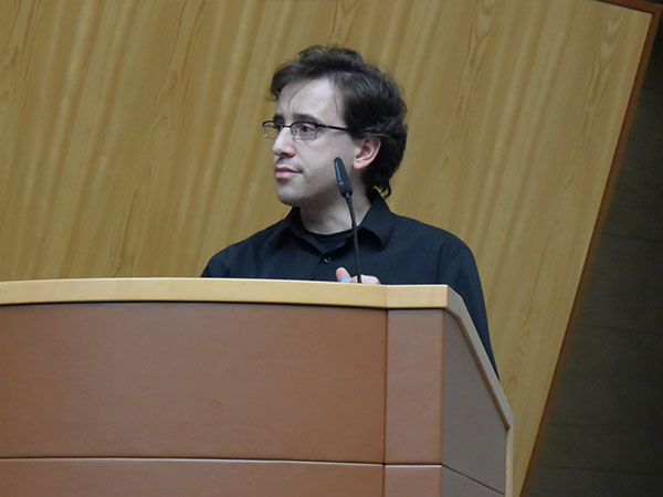
João Pereira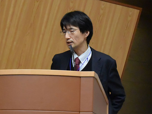
Takahshi Nagasawa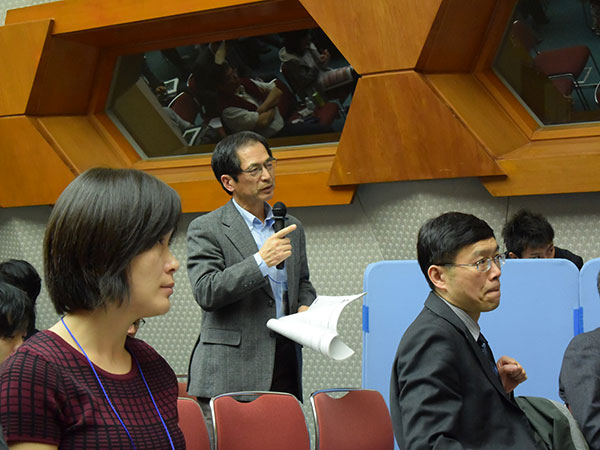
audience
Session 6
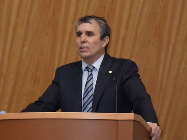
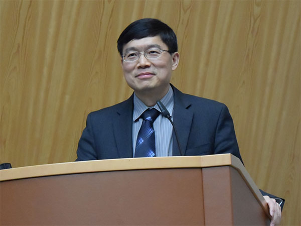
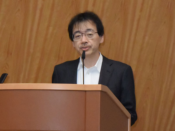
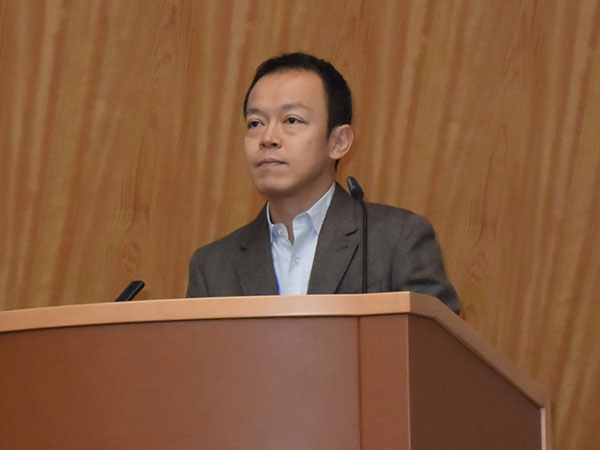
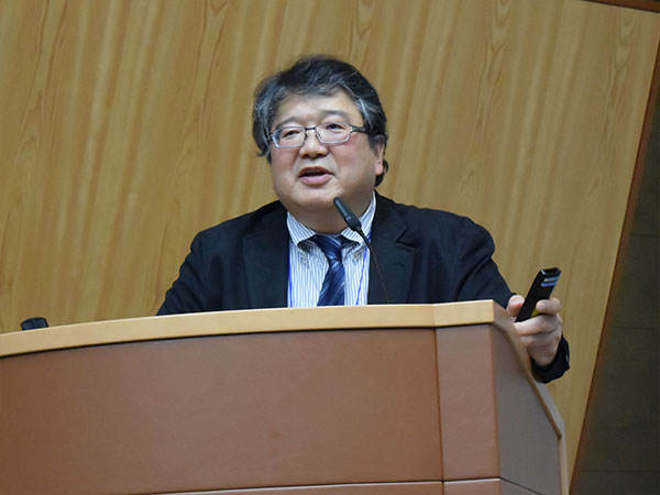
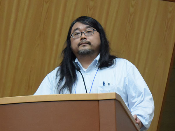
The last day started with Session 6, “Fluorescent biosensors and cell biology,” chaired by Michiyuki Matsuda, who overviewed recent progress in the biosensors based on Förster resonance energy transfer (FRET). Using cell lines and transgenic mice that expressed the FRET biosensor for ERK, he showed the similarities and the differences in the mode of growth signal transmission among epithelial cells in vitro and in vivo. Won Do Heo introduced a number of tools that could perturb cell signaling within the cells. A technology named LARIAT has been shown to inactivate molecules of interest by trapping them in specific subcellular compartments. Michael Lin talked about a novel voltage sensor named ASAP, which is the fastest optical sensor for monitoring neuronal activity. Takeaki Ozawa developed various optogenetic tools such as myr-Akt, which can mimic PI3-kinase activation. Here, fluorescent molecules and luminescent molecules were shown to be excellent tools for the development of techniques to visualize and manipulate cellular functions.
Michiyuki Matsuda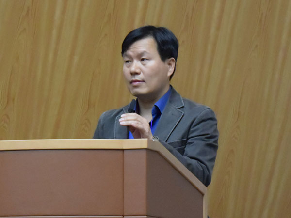
Won Do Heo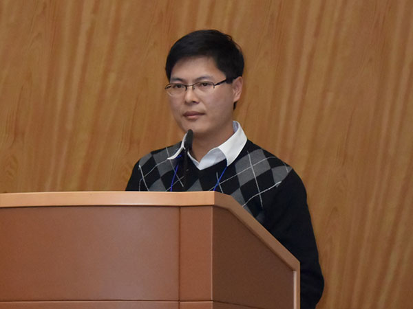
Michael Lin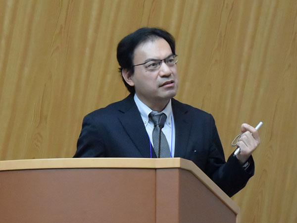
Takeaki Ozawa
Session 7
The last session, Session 7, “Fluorescence Imaging of Cardiovascular Systems,” was chaired by Shigetomo Fukuhara. Both Shigetomo Fukuhara and Suk-Won Jin discussed the mechanism of vascularization by using fluorescence imaging of zebrafish. Because of their rich mutant resources and transparency, zebrafish would seem to be the ideal system for elucidating the development of vascular systems. Seiji Takashima used ATeam, an ATP FRET biosensor, to show the correlation between oxygen supply and ATP concentration in the living heart tissue. The last speaker, Satoshi Nishimura, exhibited a number of beautiful video images covering processes ranging from thrombus formation to inflammatory cell infiltration into lipid tissues. He showed how powerfully live imaging contributes to attempts to decipher the complex mechanisms underlying metabolic syndromes.

Shigetomo Fukuhara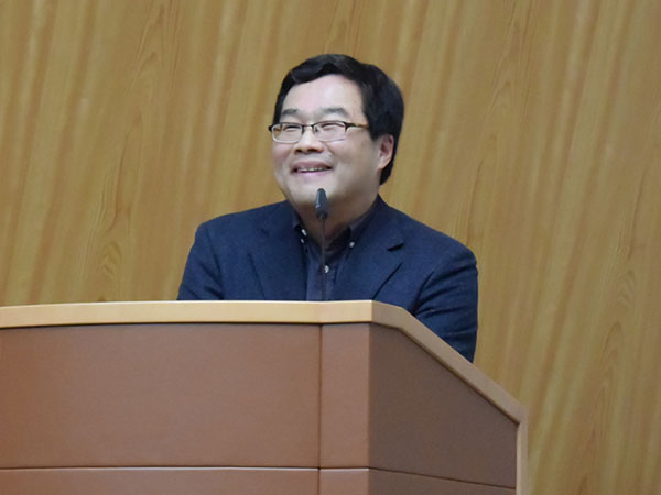
Suk-Won Jin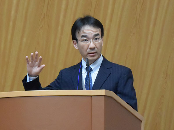
Seiji Takashima
Satoshi Nishimura
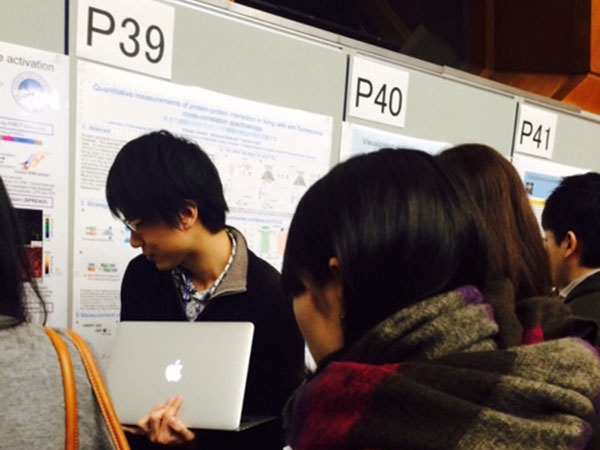
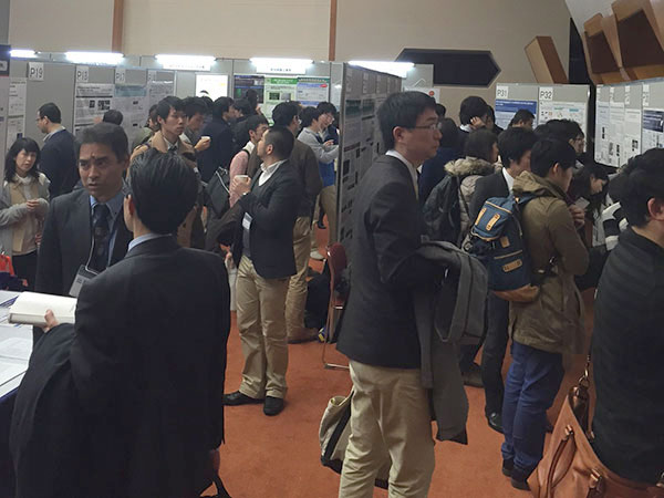
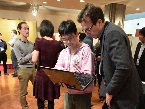
Poster sessions
The poster sessions presented more than 30 researchers and were open on the first and second days. The room was full of heated discussion. Eric Betzig was so kind as to pose in photographs with the younger researchers, who of course held him in great esteem. Guests from abroad eagerly talked to graduate students and postdocs, which was very encouraging for these young Japanese researchers.
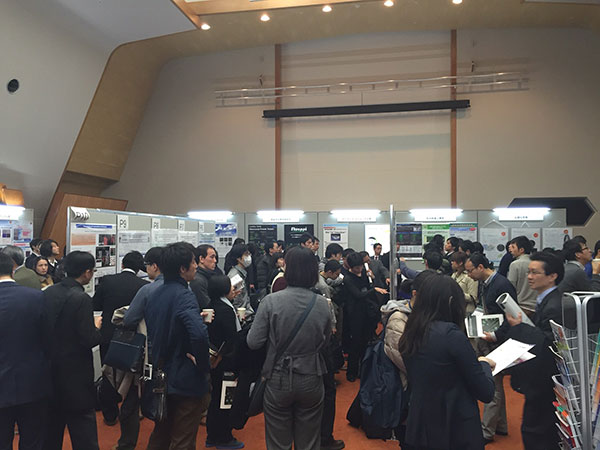
Poster sessions
Luncheon seminars
Fifteen companies exhibited their products in the room where the poster sessions were held. Needless-to-say, the providers of microscopes and other reagents are important collaborators in the promotion of fluorescent live imaging. This communication between the vendors and the researchers must have been quite important for both sides. It should also be noted that the Olympus Company presented two luncheon seminars, in which Yasushi Okada, Hideki Taniguchi, and Mototsugu Eiraku presented their remarkable cutting-edge imaging technologies.
Yasushi Okada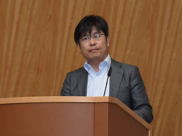
Hideki Taniguchi
Mototsugu Eiraku
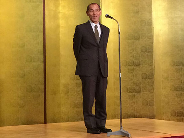
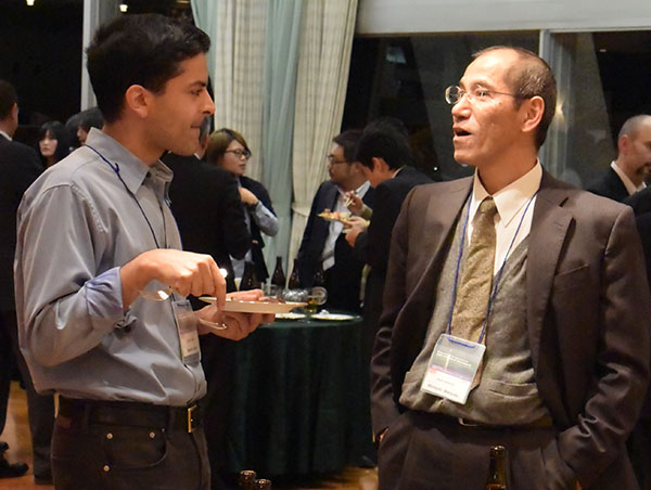
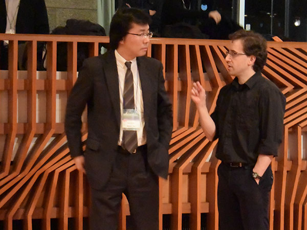
Party
During this three-day symposium, we seemed to be fully immersed in almost every conceivable area of fluorescence imaging. And indeed, as many speakers mentioned, interdisciplinary collaboration among biology, physics, and informatics will be needed for the future progress of fluorescence imaging. This symposium was intended to provide the possibility of such interactions. It appears to have worked, since many researchers could already be heard discussing potential collaborations during the coffee breaks and receptions.


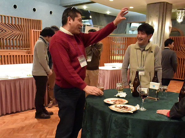
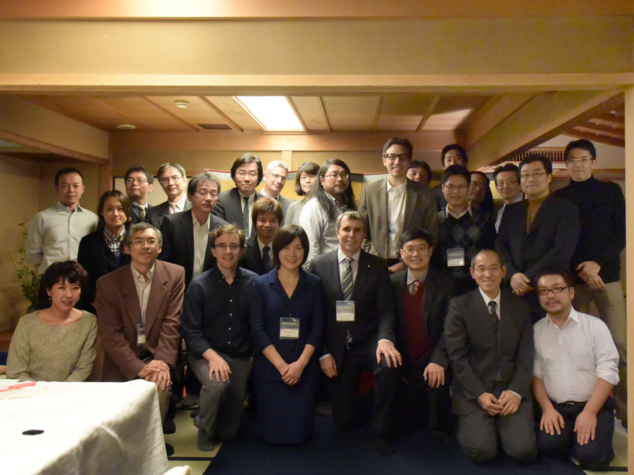
guest party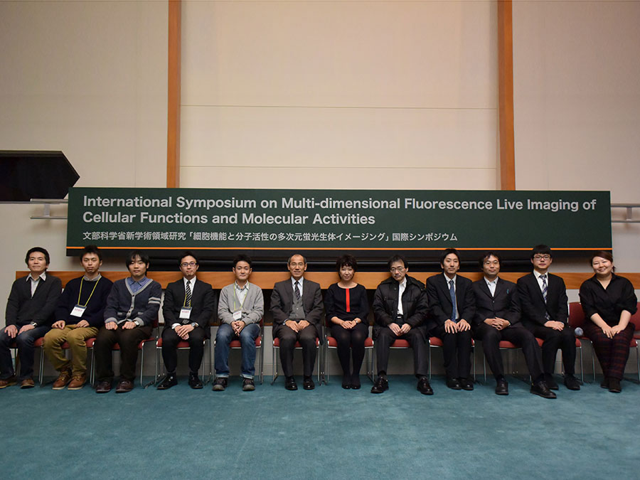
members of
Symposium Office
The research grant on “Multi-dimensional Fluorescence Live Imaging of Cellular Function and Molecular Activities” ends this March, but we are sure that the seeds planted by this meeting will continue to send forth sprouts in the near future.
Lastly, we cordially thank Kyoto University and the following foundations for their support.
The Kyoto University Foundation
The Mochida Memorial Foundation for Medical and Pharmaceutical Research
The Terumo Life Science Foundation
The NOVARTIS Foundation (Japan) for the Promotion of Science
The Naito Foundation
Written by Michiyuki Matsuda.